
Search

Sign In Join Free

1 / 1
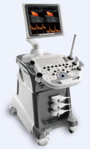
4d Color Doppler Ultrasound Scanner
Get Latest Price
Send Inquiry
| Model No. : | JAC 4D color doppler |
|---|
Longfian Scitech Co., Ltd.
You might also like


Product description
Product Description
Detailed Product Description
1. CE, ISO &FDA certificate
2.3D & 4D Ultrasound
3. Color doppler.
Image Modes
B-Mode, M-Mode, Color Doppler (CFM), Doppler Spectrum Mode (PWD), Power Doppler Mode
Measurements & Calculations
1) Basic B-mode: Distance, area and circumference, A/B ratio, angle, ellipse, volume
2) Basic M-mode: Velocity, heart rate, distance, slope, A/B ratio, time
3) Basic Doppler: Peak velocity/frequency, time, mean velocity, heart rate, RI, PI, S/D ratio, flow volume, area
4) Basic Obstetrical Package: Measurements include CRL, BPD, OFD, HC, TAD, AC, FL, APAD, FHR, user-defined measurement tables; Calculations include: EFW, GA, EDD, HC/AC, FL/AC, FL/BPD, CI; Reports cover: Measurements averaging, tables and formulas, qualitative descriptions and pictogram display
5) Basic Gynecology Package: GYN and fertility protocol, size and volume of Ovaries, Uterus and Follicles, Cervix, Endometrial
6) Basic Cardiac Package: Measurements include LVID, LVOT, LVPW, LA, LVET, LVPG for left ventricle and RVOT, RVD, RVET for right ventricle, cardiac output, cardiac index, FS, EF, heart rate; Reports cover B-mode, M-mode and Doppler mode
7) Basic Urology Package: Measurements include the volume of prostate, stepwise and residual Urine; Reports cover: PSA/PSAD entry and calculation, qualitative descriptions and pictogram display
IMAGE FEATURES
1) Display
Display Depth: Up to 28 cm,
Probe dependent: Convex: 18 steps, Linear: 14 steps
Display Gray Levels: 256; Continuous variable contrast and frame rate of up to 200 frames/sec
Display Format: B, 2B, 4B, M, B/D, B/M, B/C, B/C/MC, B/D, B/P, B/C/D
Image Orientation: Left/right B mode reversal and up/down image invert; 90, 1800 rotation
Magnification: Zoom with pan capability in real-time or freeze B, C and M mode
Annotation: Allows the user to annotate anywhere on the image with pre-defined annotation list and anatomical body marks
Screen Display: Display of all patient and exam related imaging parameters, and on-screen documentation of image parameters in single / dual display modes
2)3D & 4D Ultrasound
Offer freehand 3-D ultrasound with user training tools
Offer 3-D viewing and editing slices to remove unwanted tissue structure from arbitrary angles
Offer 3-D display in B-mode and Color Doppler simultaneously
4-D Picture forming capability
3) Image Review
CINE Review: Variable speed motion review and frame-by-frame review Storage only limited by internal memory of the system board
Standard: 1, 024 B-Mode frame and 170 seconds M-Mode data
Standard: 520 color frames and 380 seconds Doppler Spectrum
Post Procession and measurements
4) Image Management
Storage only limited by hard disk of the system board
Standard: 320 GB hard disk drive for local image storing
CD-RW/DVD Drive as removable read, write, archive and storage, USB, S-Video, VGA
Standard: 2, 000 Images w/ TIF format on CD
DICOM & PACs Compatible
Standard Configuration
1) Main unit, 17"LCD monitor, 3 probe connectors, 2 USB ports
2)3.5MHz Convex Array Probe,
3)PW, PWD, PWR
Optional
Linear Probe: 7.5MHz
Trans-vaginal: 7.5 MHz
Phased array: 2.5 MHz
Micro convex: 5.0MHz
4D Transducer: Curved 3.5 MHz
Video printer
3D software
DICOM, CW, Free Steering M,
Send your inquiry to this supplier
Send Inquiry
Product Alert
Subscribe to your interested keywords. We will send freely the latest and hottest products to your Inbox. Don't miss any trade information.
Subscribe
Your use of this website constitutes acknowledgement and acceptance of our Terms & Conditions.
Copyright © 2009-2024 Bossgoo Co., Ltd. All rights reserved.
